 Sungjoon Park
2025.12.03
Sungjoon Park
2025.12.03
[CTAD 2025] MOIRA, a multimodal framework for more effective prediction of Alzheimer's disease with incomplete data
Introduction
AI-based disease prediction models are demonstrating performance comparable to or surpassing that of physicians or pathologists in certain areas[1]. Particularly for complex brain disorders like Alzheimer's disease, a multimodal approach integrating diverse information such as cognitive assessments, genetic data, MRI scans, and blood biomarkers is essential. However, fundamental challenges must be addressed before these technologies can be applied in actual clinical settings or clinical trials.
At this year's CTAD (Clinical Trials on Alzheimer's Disease) 2025 conference, LG AI Research's presentation addresses the challenges AI must overcome to move beyond the laboratory environment and contribute to actual patient care. Pathological changes in the brain begin 10 to 16 years before Alzheimer's symptoms appear[2]. Therefore, early detection is key for preventative intervention, which requires the effective utilization of as much patient data as possible.
Limitations of multimodal biomedical data: Incomplete modality
The data available for use in the bio domain, particularly in clinical research, is extremely diverse. From multi-omics data such as genomics, transcriptomics, and proteomics, to magnetic resonance imaging (MRI), cognitive assessment scores, cerebrospinal fluid (CSF) biomarkers, and clinical history, each modality contains information about unique dimensions of disease.Integrating these unstructured data or time-series data can provide a holistic understanding of disease progression.
However, clinical data has an inherent limitation: the issue of incomplete modality. In real clinical settings, it is practically impossible for every patient to undergo every type of test without omission. As observed in the public data we used in our research, due to differences in protocols, cost constraints, limitations in biopsy specimens, and the unique nature of research objectives, many patients in the dataset have only partial modalities. Taking Alzheimer's as an example, MRI scans, which provide direct clues for diagnosis, are often not performed until significant symptoms appear due to cost issues. Similarly, CSF biomarkers represent significant indicators of Alzheimer's disease, but they also are fundamentally limited due to being invasive tests. Furthermore, when data or testing standards change, existing models may no longer be able to process new data.

Image 1. Number of samples per modality combination in the Alzheimer's public datasets ROSMAP[3] and ADNI[4]
However, existing multimodal AI research has not sufficiently reflected this reality, relying solely on complete datasets where every sample possesses all features to develop predictive models. As a result, these models were developed using a limited set of modalities, and some patients' data were excluded from the research due to missing modalities. This not only reduces the utility of the dataset but also impairs the model's predictive accuracy and its generalized performance in responding to various clinical situations.
Relatively recent studies have proposed attempts to handle data with incomplete modalities, but they showed limitations where applying the same model to more data resulted in decreased clinical classification accuracy. Particularly, some multimodal prediction models failed to effectively utilize the rich information from imaging data such as MRI scans, which directly reflects neurodegeneration.
MOIRA, generating embeddings from incomplete modalities
We have developed a methodology that maintains high predictive accuracy without excluding incomplete data. MOIRA (Multi-Omics Integration with Robustness to Absent modalities)[3] is a multimodal integration framework that provides reliable predictions even when modalities are missing. The core principle of MOIRA's operation is to transform heterogeneous clinical data into latent representations and project them into a shared embedding space. The MOIRA framework consists of three core components: encoders, an adaptive fusion module, and a predictor.
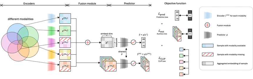
Image 2. Structure of MOIRA. Encoders, fusion module, predictor module[3]
First, dedicated encoders for each modality map data from different modalities into an embedding space of the same dimension. The key point here is that any form of data is ultimately represented as a vector of the same dimension. Next, the adaptive fusion module constructs an integrated embedding using only the available modalities. This module introduces learnable weight values for each modality and masks out missing modalities, setting their contribution to zero. It then redistributes weights only among the modalities for which data exists via softmax, and generates the integrated embedding through weighted summation. Finally, the predictor module takes this integrated embedding as input and performs the final classification.
During training, three loss functions are combined for optimization: (1) a prediction loss for the integrated embedding, (2) auxiliary loss functions for each modality's embedding, and (3) a CLIP-style contrastive loss function. This enables individual modality embeddings to harbor discriminative power, aligns modality embeddings with the integrated embedding within the same representation space, and prevents modality collapse.
The advantage of this design is that it can utilize data regardless of the combination of modalities a patient is available of. During the training phase, it can incorporate a larger number of patient samples, and during the inference phase, it can provide predictions for patients who have not undergone all examinations. Furthermore, this framework operates with arbitrary encoders and prediction modules. In fact, our experiments showed that processing raw MRI data with a 3D CNN-based encoder yielded superior performance compared to using preprocessed features summarized using the FreeSurfer software[4], demonstrating the successful integration of an encoder that preserves spatial and morphological structural information into the MOIRA framework.
To validate our approach, we tested two datasets that are widely benchmarked in Alzheimer's research. The first dataset is ROSMAP[5], which includes five types of multi-omics data extracted from postmortem brain tissue, and among the total 748 samples, only 11% (86 samples) include all five modalities. The second dataset is ADNI[6], a longitudinal study that collected clinical records, MRI scans, biomarkers, and genetic information from patients. Among the total 2,380 samples, only 56% (1,323 individuals) are available of the complete set of modalities. If only complete data were used, both datasets would inevitably suffer significant loss of information.
Optimizing dataset utilization, prediction accuracy, and clinical applicability
Experimental results show that MOIRA achieved an accuracy of 0.920 on ROSMAP and 0.911 on ADNI. This represents state-of-the-art performance compared to previous research using these datasets. Especially in our study using the ADNI dataset, we observed a trade-off from prior studies, where as the number of utilized samples increases, the prediction accuracy drops. MOIRA breaks out of this trade-off, improving both improving the data coverage and high prediction at the same time. We also quantified the contribution of each modality through ablation studies on the ROSMAP dataset and found that utilizing all modalities and additional samples thereof is beneficial for improving performance. In particular, when using the same data as previous studies (Trimodal (∩) in the lower left of image 3), the predictive performance was no better than that of previous studies. This implies that MOIRA's high predictive accuracy stems not from a unique model architecture, but from dataset expansion and effective integration.
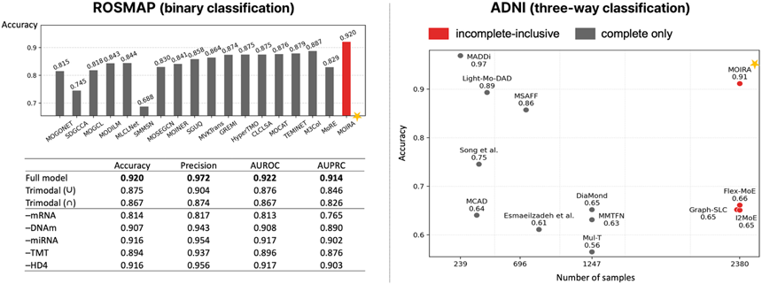
Image 3. Prediction accuracy evaluated on the ROSMAP and ADNI datasets and ablation studies of ROSMAP[3]
As mentioned earlier, it is difficult for patients to undergo all tests in actual clinical practice. MOIRA can offer its best prediction using only a subset of the test results of which the patients are available. Notably, we had an interesting observation regarding the role of MRI data in the ADNI experiment. An experiment was conducted where MRI was included during training but excluded during inference. We conducted an experiment in which MRI data was included during training but excluded at inference. Compared to the setting where MRI data was unavailable for both training and inference, the MRI-informed model showed an approximately 12-percentage-point improvement in accuracy (0.693 vs. 0.619). This suggests that MRI data can facilitate the learning of representations from the other modalities. The case provides a strong example that, when incomplete modalities are properly utilized, the integrative use of multiple modalities has its greatest potential. Such findings can also contribute to improving real-world healthcare systems. If a model can be developed to detect risk early using only blood tests or cognitive assessments, without the need for relatively costly MRI scans, a diagnostic strategy that recommends MRI scans only for patients likely to have Alzheimer’s becomes feasible. This would enable more cost-effective use of medical resources and facilitate earlier diagnosis.
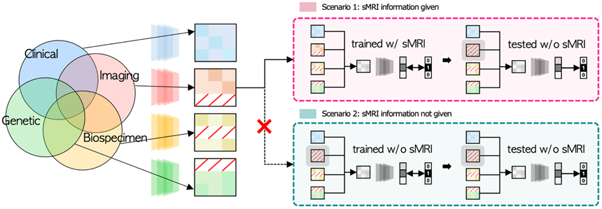
Image 4. Design of experiment to assess the impact of sMRI (structural MRI) data[4]
For clinical deployment of an AI model, rigorous biological validation is required in addition to high predictive performance. In our study, we used Integrated Gradients to select features that significantly contributed to the model's predictions. We identified the top 12 features for each omics type in the ROSMAP dataset, and of the 60 features thus identified, 57 were previously reported in existing Alzheimer's disease research literature. For example, these included SCD and KIF5A in mRNA, TMEM59 in DNA methylation, miR-132 in microRNA, GFAP in proteomics, and alpha-GPC and myo-inositol in metabolomics. These are all molecules known to be involved in Alzheimer's disease pathology. This demonstrates that MOIRA captures signals actually reflecting the disease's biological mechanisms, rather than merely learning statistical patterns.
Conclusion
LG AI Research plans to expand follow-up research in the following directions, with a focus on discovering meaningful biomarkers using MOIRA and applying it to clinical diagnostics. For the practical clinical application of our research, intensive efforts are required in the following subsequent research directions. In line with our findings, a natural direction is to design modality-specific encoders, akin to the 3D CNNs used for MRI data, to further enhance predictive utility and to strengthen explainability so that medical professionals can better understand the model’s reasoning. Furthermore, exploring the mechanism by which MRI enhances representation training across other modalities, as shown above, may capture interactions between multiple modalities and lead to new discoveries.
The moment AI technology truly demonstrates its value is when it transcends laboratory benchmarks to create positive change in people's lives. Through our presentation at CTAD 2025, we proposed an effective methodology for addressing the incomplete modality problem in the biomedical domain and demonstrated its utility. By leveraging incomplete modalities, we can fully utilize datasets, leading to improved prediction model accuracy and enhanced practical applicability. This approach is expected to be widely applicable not only to Alzheimer's-related research, but also to general fields where incomplete modalities exist.
- 참고
[1] Athanasopoulou, Konstantina, et al. "Artificial intelligence: the milestone in modern biomedical research." BioMedInformatics 2.4 (2022): 727-744.
[2] Greenberg, Barry D., et al. "CSF Alzheimer disease biomarkers: time-varying relationships with MCI symptom onset and associations with age, sex, and ApoE4." Neurology 99.15 (2022): e1640-e1650.
[3] Park, Sungjoon, et al. "Robust Multi-Omics Integration from Incomplete Modalities Significantly Improves Prediction of Alzheimer's Disease." Proceedings of the ICML 2025 Workshop on Multi-modal Foundation Models and Large Language Models for Life Sciences.
[4] Lee, Juhyun, et al. "Structural MRI?Informed Multimodal Fusion for Robust Alzheimer’s Disease Prediction." NeurIPS 2025 Workshop for Imageomics: Discovering Biological Knowledge from Images Using AI.
[5] Perez-Gonzalez, Alejandra P., et al. "The ROSMAP project: aging and neurodegenerative diseases through omic sciences." Frontiers in Neuroinformatics 18 (2024): 1443865.
[6] Petersen, Ronald Carl, et al. "Alzheimer's disease Neuroimaging Initiative (ADNI) clinical characterization." Neurology 74.3 (2010): 201-209.