 Minsoo Lee
2026.04.21
Minsoo Lee
2026.04.21
 Jonghyun Kim
2026.04.21
Jonghyun Kim
2026.04.21
[AACR 2026] Spatial biology and AI research at LG AI Research-VUMC
3D spatial biology, pathological AI, and multi-omics foundation models
The importance of spatial information in cancer research has been increasing, as tumors are not merely collections of abnormal cells, but rather complex microenvironments in which tumor cells, immune cells, and stromal cells formed specific structures and interactions. Consequently, recent research has moved beyond simply examining gene or protein expression levels to interpreting where these signals were located within tissues, the structures in which they appeared, and how they changed over time.
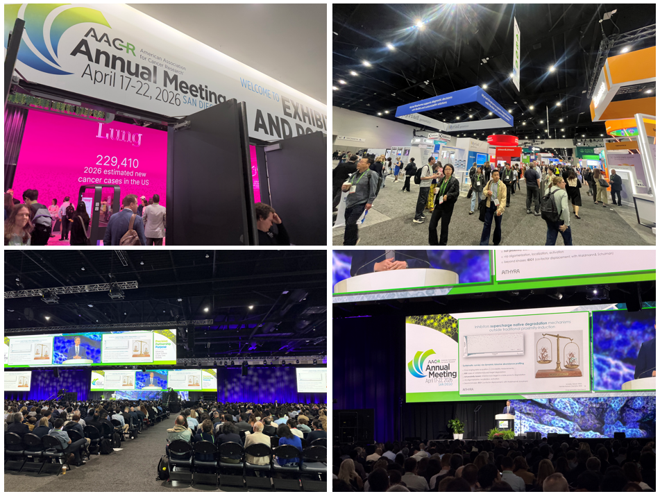
Image 1. Scene from 2026 AACR
In line with this trend, LG AI Research and Vanderbilt University Medical Center(VUMC) have conducted various joint research projects that integrated spatial biology with AI. At the 2026 AACR, we shared several studies spanning spatial biology, 3D/4D image analysis, experimental design for spatial analysis, pathology AI, and foundation models. These studies were originated from distinct scientific questions. Some focused on more precisely characterizing the three-dimensional spatial context of gastric cancer tissue, while others aimed to track the dynamic processes of drug delivery and intracellular activity. The research also expanded to include studies concentrated on optimizing the selection of regions of interest (ROIs) in spatial analysis experiments and extending the integration of pathological images and molecular signals into a larger-scale representation learning frameworks.
Through collaborative research with Bio Intelligence lab at LG AI Research and VUMC has developed a range of AI models aimed at advancing cancer research. In this post, we highlight several of the key studies presented at the 2026 AACR, organized around major research themes.
1. Visualizing Cancer Tissues through 3D Spatial Multi-omics
Spatial transcriptomics and spatial proteomics are powerful tools for understanding the molecular architecture within tissues. However, as the actual tumor microenvironment inherently has a three-dimensional structure, it is often difficult to fully grasp tissue continuity and structural context from a single tissue section. This limitation is particularly evident in gastric cancer, which have complex glandular structures and intricate interactions between epithelial cells, stromal cells, and immune cells.
In 3D Multimodal Spatial Profiling of Pre-Gastric Cancer Progression Using Same-Slide RNA–Protein and Fluorescent H&E, we aimed to interpret the epithelium, stroma, and immune niches surrounding gastric cancer lesions more precisely by combining 3D holotomography with RNA, protein, and fluorescent H&E data measured on the same slide. A key aspect of this study is that the RNA, protein, and morphological data are not derived from different samples, but rather constitute multimodal data aligned on the same slide coordinates. This allows us to directly link morphological and molecular features within the same tissue context and to re-interpret, within a three-dimensional context, whether lesions observed in two dimensions are actually continuous or discrete structures.
Three-Dimensional Spatial Multi-omics of Gastric Tumor Sections Reveals Hidden Features Beyond 2D Analyses investigates immune niches and tumor microenvironment structures that are difficult to capture through the analysis of a single 2D section, by performing 3D reconstruction on a series of consecutive sections. By aligning multiple serial sections within a common coordinate system and analyzing RNA, proteins, and holotomography together, we sought to determine whether signals that appeared weak in two dimensions could emerge as more distinct spatial regions in three dimensions.
These two studies are significant in that they point toward a shift from two-dimensional analysis to a more spatially integrated, three-dimensional interpretation of the tissue architecture. They demonstrate that reinterpreting these signals within the three-dimensional continuity of tissue is becoming just as important as increasing the resolution of spatial multi-omics.
2. Beyond 3D to 4D: Tracking the dynamic processes of drug delivery and action
Going a step beyond simply observing structures leads to the challenge of interpreting biological processes such as drug delivery and action along a temporal axis. In particular, for therapeutics like ADCs, where cellular uptake, lysosomal processing, and drug release occur sequentially, it is difficult to fully explain their mechanisms of action using only single-time-point images.
In AI-driven live 3D/4D spatial multimodal evaluation of a HER2-targeting ADC: delivery, lysosomal cleavage, payload action, and bystander effects resolved by holotomography and fluorescence, we analyzed membrane binding, intracellular migration, lysosome delivery, drug action, and ripple effects over time for Enhertu, a HER2-targeted ADC, by combining real-time 3D and 4D holotomography with fluorescence signals.
In this study, we quantitatively analyzed how the drug traverse intracellular compartments, the temporal dynamics of their journey to lysosomes, and the migration patterns observed at the organoid level, using AI-based organelle segmentation and fluorescence signal alignment. Furthermore, by integrating real-time imaging results with same-slide RNA and protein profiling and spatial laser dissection, we identified niches where delivery efficiency or drug responsiveness differed.
What makes this study interesting is that it does not view drug action merely as a simple outcome, but rather seeks to observe it as a series of sequential stages: delivery, processing, manifestation of pharmacological effects, and side effects. This serves as an example of how spatial biology is expanding beyond static structural analysis to interpret the therapeutic process itself in spatiotemporal terms.
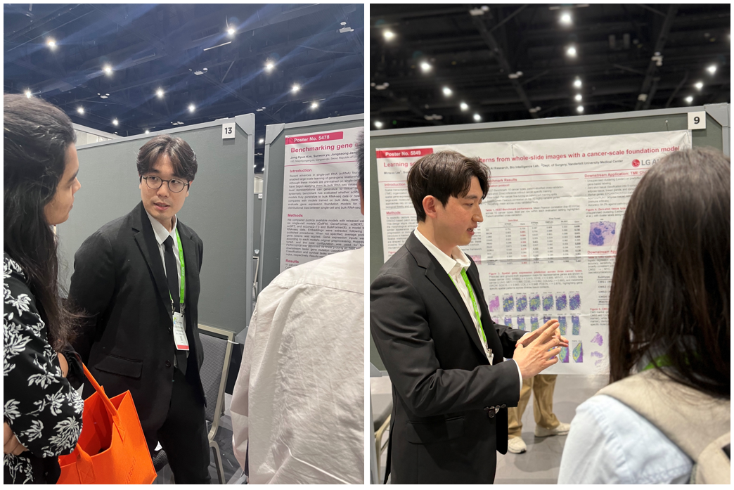
Image 2. Poster session presentation
(Left)Jonghyun Kim-Benchmarking gene expression foundation models on bulk RNA-Seq data,
(Right)Minsoo Lee-Learning spatial transcriptomic patterns from whole-slide images with a cancer-scale foundation model
3. Analytical interfaces and predictive models for better interpretation of complex spatial data
As spatial multi-omics and 3D imaging technologies advance, the challenge of transforming data into interpretable forms has become just as important as generating the data itself. This is because researchers need to explore, annotate, and discuss complex 3D data in practice.
AI-Augmented Immersive 3D and 4D Spatial Analysis Interface for Cancer Research is an interface study designed to address these challenges. In this study, we developed an immersive analysis environment that enables users to explore 3D data from cells, organoids, and tissues in real time, as well as perform segmentation, measurement, and annotation tasks within virtual and augmented reality environment. We integrated a conversational interface based on our in-house AI agent, ‘ChatEXAONE’, allowing users to access literature context and interpretive guidance during analysis. This research is significant in that it goes beyond simply visualizing complex spatial data; it represents an attempt to expand the analytical environment into one where researchers can actually view and interpret data together.
ProteoBridge: Bridging Skipped Sections via Histology-Based Protein Prediction addresses the practical limitation of being unable to directly measure proteins across all serial sections. This study aims to enable denser 3D protein reconstruction by predicting protein maps for unmeasured sections based on H&E, protein, and RNA data obtained from measured sections. A key feature of this study is the use of both H&E morphological information and transcriptomic context to reconstruct protein maps, supported by experimental results from an H&E-based protein prediction model.
Although the two studies take different approaches, they both address the challenge of transforming spatial data into a more interpretable and actionable forms. One study focuses on changing the way researchers interact with data through an analytical interface, while the other proposes a method for filling in gaps in unmeasured sections using predictive models.
4. How to optimize the selection of the region of interest in a spatial analysis experiment
Although spatial transcriptomics and multi-omics analyses provide a wealth of information, they are limited in the number and size of regions of interest. Consequently, the biological insights ultimately obtained can vary significantly depending on which regions are selected. For this reason, the selection of regions of interest should be viewed not merely as a preprocessing step, but as a critical design decision that determines the informational efficiency of the experiment itself.
Image-Based ROI Selection for Spatial Transcriptomic Experiments Using Immune Checkpoint Inhibitor (ICI) Treatment Outcome Prediction in Gastric Cancer (GC) describes a method for predicting immune checkpoint inhibitor response patterns in whole-slide H&E images of gastric cancer and, based on these results, selecting regions of interest for spatial transcriptomic analysis. The key feature is that it does not simply select areas with high tumor density, but rather proposes regions of interest that specifically reflect predicted response areas, predicted non-response areas, and areas exhibiting mixed patterns.
In this study, we first identified valid tile-level regions using cell-type and tumor classifiers based on ‘EXAONE Path’, LG AI Research’s pathology foundation model. We then calculated tile-specific scores using a weakly supervised response prediction model. We then calculate candidate regions of interest by taking into account the capture window size and rotation of the actual spatial analysis platform, thereby linking image-based predictive signals to actual experimental designs.
This study is significant in that it expands the scope of spatial omics beyond merely interpreting measurements after they are taken, to include the question of what should be measured first.
5. Research on foundation models that link pathological images and molecular signals
Among the studies shared at 2026 AACR are addressing several large-scale pathological AI and multi-omics representation learning. These studies go beyond the detailed analysis of individual experimental systems and focus on learning the connections between pathology images and molecular information in a more generalizable form.
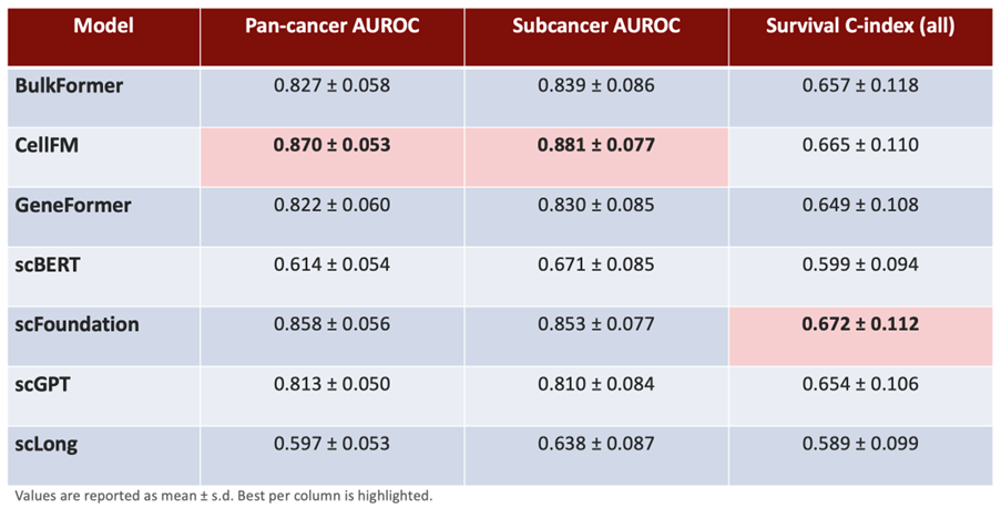
Image 3. Summary of downstream benchmark performance across foundation models.
In Benchmarking gene expression foundation models on bulk RNA-Seq data, we systematically compare the generalizability of gene expression foundation models in bulk RNA-seq settings. Although many recent models have been pre-trained primarily on single-cell data, they are widely used in practical applications involving bulk RNA-seq as well. Through mutation classification and survival prediction, this study aims to determine which models demonstrate more stable transfer ability across tasks.
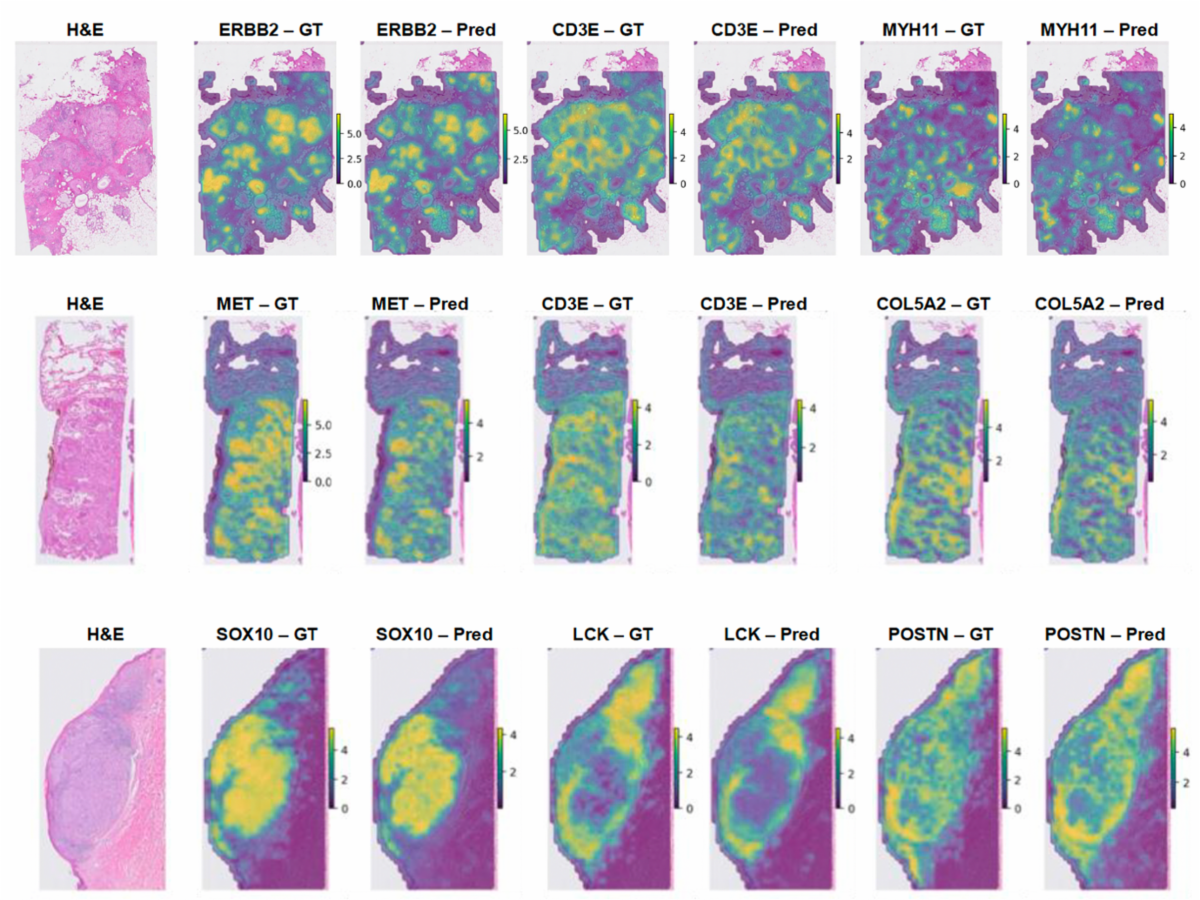
Image 4. Spatial gene expression prediction across three cancer types.
Predicted and ground-truth expression maps for representative genes are shown in breast cancer
Learning spatial transcriptomic patterns from whole-slide images with a cancer-scale foundation model addresses the challenge of directly predicting spatial transcriptomic patterns from standard H&E whole-slide images. By co-training a DINOv2-based patch encoder with a spatial transcriptomic prediction module and utilizing a masked transformer that integrates contextual information from neighboring patches, we aimed to incorporate tumor microenvironment signals, which cannot be explained by local morphology alone, into the model’s representations.
Omics-aware patch aggregation via multimodal co-training with a scalable multi-omics encoder for slide-level prediction across an oncology biomarker panel is a framework that jointly trains on pathology images and multi-omics data using a contrastive learning approach. Specifically, to reflect real-world clinical settings in which not all data are fully paired, we proposed a multimodal learning architecture based on partially paired data. Through this approach, we aimed to address a broader range of predictive tasks, including expression-based biomarkers, mutations, MSI, and TMB.
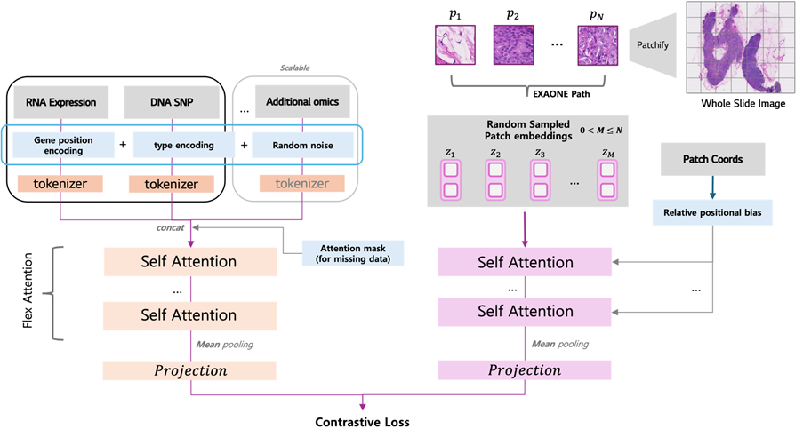
Image 5. Omics-Aware Patch Aggregation Framework. The Slide Encoder (SE) processes WSI patches with coordinate-aware attention, while the Multi-Omics Encoder (MOE) jointly models RNA and DNA modalities via a shared Transformer. Both are aligned through a slide-level contrastive objective.
While these studies do not directly address spatial biology itself, they point to an important direction in the field of representation learning that links pathology images with molecular information.
In conclusion
Ahead of AACR 2026, LG AI Research and VUMC collaborated on a variety range of topics , including 3D spatial multi-omics, real-time 3D and 4D drug analysis, immersive analysis interfaces, optimization of region-of-interest selection, pathology foundation models, and multimodal learning across omics data.
Rather than pushing a single, overarching narrative, these studies highlight several key research topics at the intersection of spatial biology and AI. These topics collectively reflect current research trends aimed at observing cancer tissues with greater precision, interpreting complex data more effectively, and extending these insights to address molecular and clinical questions.
We hope that these studies shared at the AACR 2026 will serve as a meaningful starting point for broader discussion on the next stage of AI-driven cancer research and spatial biology.
| Research papers | Key details |
| 3D Multimodal Spatial Profiling of Pre-Gastric Cancer Progression Using Same-Slide RNA–Protein and Fluorescent H&E | By integrating RNA, protein, and fluorescent H&E staining on a single slide and combining this with 3D holotomography, we three-dimensionally reconstructed the epithelium, stroma, and immune niche of precancerous gastric lesions. This approach identifies the spatial structures associated with lesion progression, which were difficult to capture using conventional 2D analysis, at a three-dimensional level. |
| AI-Augmented Immersive 3D and 4D Spatial Analysis Interface for Cancer Research | An immersive analysis platform combining an AI chatbot (chatEXAONE) with a VR/AR environment. It allows real-time exploration of volume data of cancer cells, organoids, and tissues, and intuitively visualizes the tumor microenvironment and drug delivery processes. |
| AI-driven live 3D/4D spatial multimodal evaluation of a HER2-targeting ADC: delivery, lysosomal cleavage, payload action, and bystander effects resolved by holotomography and fluorescence | The intracellular journey of Enhertu, an HER2-targeted ADC, was tracked using AI-based 3D/4D live imaging. The process was elucidated in real time, from lysosomal cleavage of the GGFG linker to the release of DXd and the bystander effect, and this presents an integrated pipeline that captures delivery inhibition and resistance niches by combining these findings with molecular profiling of fixed tissues and spatial laser sorting. |
| Image-Based ROI Selection for Spatial Transcriptomic Experiments Using Immune Checkpoint Inhibitor (ICI) Treatment Outcome Prediction in Gastric Cancer (GC) | A strategy for automatically selecting ROIs based on ICI treatment response predictions in H&E images. By moving beyond simple tumor region selection and spatially reflecting predicted response, non-response, and mixed patterns, it can efficiently capture niches directly linked to treatment outcomes. |
| ProteoBridge: Bridging Skipped Sections via Histology-Based Protein Prediction | A deep learning framework that learns multimodal information on H&E, proteins, and RNA to predict protein expression across entire sequences. It compensates for skipped fragments to densely reconstruct 3D tumor protein maps and fills data-driven gaps in protein profiling between fragments. |
| Three-Dimensional Spatial Multi-omics of Gastric Tumor Sections Reveals Hidden Features Beyond 2D Analyses | By integrating RNA, protein, and holotomography data in the x–y–z dimensions, it reveals immune niches and TME structures that were not visible in single-slide analysis. It experimentally demonstrates the limitations of 2D analysis and presents a practical strategy for 3D tumor-immune profiling. |
| Benchmarking gene expression foundation models on bulk RNA-Seq data | A benchmarking study that systematically compares and evaluates various RNA foundation models based on bulk RNA-seq data. |
| Learning spatial transcriptomic patterns from whole-slide images with a cancer-scale foundation model | Development and benchmarking of a cancer-scale foundation model that predicts gene expression levels by learning spatial transcriptomic patterns from H&E WSI. |
| Omics-aware patch aggregation via multimodal co-training with a scalable multi-omics encoder for slide-level prediction across an oncology biomarker panel | A patch aggregation methodology that combines EXAONE Path and omics information through multimodal collaborative learning. Benchmarks slide-level predictions and performance across a panel of tumor biomarkers. |